High Performance Tomographic Image Reconstruction
We exploit programmable commodity graphics hardware boards (GPUs) to accelerate a wide variety of 3D computer tomographic (CT) reconstruction algorithms for a number of application settings and energy modalities. Once a suitable parallel algorithm and implementation has been devised, we typically reach speed-ups of 1-2 orders of magnitude, without significant loss in reconstruction quality -- as is shown below.
 |
 |
 |
 |
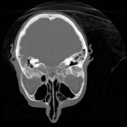
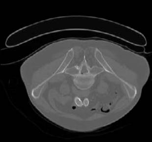
| Original | GPU reconstructed |
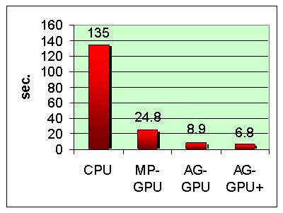
The above volumes were GPU reconstructed with the Feldkamp filtered backprojection algorithm from 360 projections into a 5123 volume (at floating point precision). The bar charts below illustrate the reconstruction performance in seconds (top) and processed projections per second (bottom). Platform: NVIDIA 8800 GTX.
 |
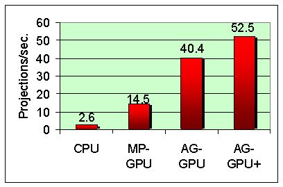 |
These charts compare the performance of a fairly optimized CPU-based reconstruction with GPU-accelerated reconstruction. Here, MP-GPU uses only the parallel GPU pipelines (fragment shaders), AG-GPU incorporates the graphics subsystem as well, and AG-GPU+ adds further optimizations using higher-level graphics pipeline constructs. While other GPU-accelerated CT reconstruction frameworks have been recently published, these tend to only operate on the MP-GPU level, with the limited performance gains shown here (acceleration with NVIDIA CUDA tend to operate at this level as well). By incorporating hardwired graphics pipelines, the performance can be boosted nearly 4-fold.
Our RapidCT system enables fast, accelerated 3D reconstruction in diagnostic imaging, radio-therapy applications, surgery planning, electron microscopy, and others, at a fraction of the cost of proprietary devices. Further applications have been the iterative 3D reconstruction from data acquired with transmission ultrasound for breast mammography, data obtained from MV-CT and proton-CT scanners for the treatment of cancer, projections obtained via mobile X-ray source/detector pairs, and others.
The Publications link in the menu bar has all publications (and more). Some of these applications are also highlighted in the Application link.
This project was funded in part by the NIH (grant # R21 EB004099-1) and the Keck Advanced Microscopy Laboratory, University of California, San Francisco.