Human Vision and Color Perception
Vision is an ability to see the features of objects we
look at, such as color, shape, size, details, depth, and contrast. Vision
is achieved when the eyes and brain work together to form pictures of
the world around us. Vision begins with light rays bouncing off the
surface of objects. These reflected light rays enter the eye and are
transformed into electrical signals. Millions of signals per second
leave the eye via the optic nerve and go to the visual area of the brain.
Brain cells then decode the signals into images, providing us with sight.
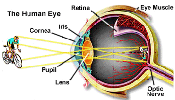
Light rays reflected from any object we look at enter the eye and are
focused by the eye's optical structures: cornea, iris, pupil, and lens.
The final destination of the light rays is the retina, a layer of nerve
tissue that lines two-thirds of the back of the eye. In the center of
the retina is the macula, an area that is only 1.5 mm (0.06 in) in diameter.
The macula is responsible for the clearest, most detailed vision. The
retina is made up of two types of cells: cones
and rodes
(click the links to find more).
Light rays that reflect from the upper half of any object we look at
are focused on the lower half of the retina. Rays from the lower half
of the same object are focused on the upper half of the retina. This
would seem to give us an upside-down picture of the world. Fortunately,
these signals are rearranged when the brain processes them into an image
that is right side up.
Another feature of eyesight is stereoscopic vision, the ability of both
eyes to look straight ahead but see the same scene from a slightly different
angle. The eyes' visual fields overlap in the center, and the brain
merges these images to create a sense of depth important for judging
distance. Humans and other mammals have stereoscopic vision. Birds,
fish, and snakes have monocular vision in which each eye sees a separate
image covering a wide area on each side of the head.
Below is a graphical illustration of the spatial arrangement of rod
and cone cells and their connection to neurons within the retina. Rod
cells have a peak sensitivity to green light (about 550-555 nanometers),
although they display a broad range of response throughout the visible
spectrum. They are the most populous visual receptor cells with each
eye containing about 130 million cells. The light sensitivity of rod
cells is about 1000 times that of cone cells, however the images generated
by rod stimulation alone are relatively unsharp and confined to shades
of gray, similar to those found in a black and white soft-focus photographic
image. Rod vision is commonly referred to as scotopic or twilight vision
because in low light levels it allows us to distinguish shapes and the
relative brightness of objects, but not their colors.
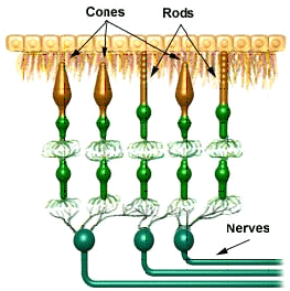
Cones consist of three types of cells each "tuned" to a distinct
wavelength maxima of response centered at either 430, 535, or 590 nanometers.
The population of cone cells is much smaller than rod cells with each
eye containing about 7 million cone cells. True color vision is induced
by the stimulation of cone cells, but the relative intensity of stimulation
of each of the three cone receptors determines the color that is imaged.
For example, a beam of light that contains mostly blue short-wavelength
radiation stimulates the cone cells that respond to 430 nanometer light
far more than the other two cone types, and we see that light as blue.
Light with a majority of wavelengths centered around 550 nanometers
is seen as green, and a beam containing mostly 600 nanometer wavelengths
or longer is seen as red. Pure cone vision is referred to as photopic
vision and is dominant at normal light levels, both indoors and out.
When all three types of cone cells are stimulated equally, we perceive
the light as being achromatic or white. As an example, noon sunlight
appears to us as white light because it contains approximately equal
amounts of red, green, and blue light. An excellent demonstration of
the color spectrum of sunlight is interception of the light by a glass
prism, which refracts (or bends) different wavelengths to varying degrees,
spreading out the light into it's component colors. Our color perception
is dependent upon the interaction of all receptor cells with light and
this combination results in nearly trichromic stimulation. There are
shifts in color sensitivity with variations in light levels so blue
colors look relatively brighter in dim light and red colors look brighter
in bright light. This effect can be observed by pointing a flashlight
onto a color print, which will result in the reds suddenly appearing
much brighter and more saturated.
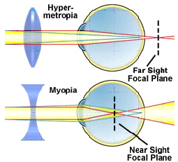
Focus in the eye is controlled by a combination of elements including
the iris, lens, cornea, and muscle tissue that can alter the shape of
the lens so the eye can focus on both nearby and distant objects. However,
in some instances these muscles do not work properly or the eye is slightly
altered in shape and the focal point does not intersect with the retina.
As people age, the lens becomes harder and cannot be focused properly
leading to poor vision. If the point of focus is short of the retina
(drawing above), the condition is called nearsightedness or myopia and
people with this affliction cannot focus on distant objects. In cases
where the focal point is behind the retina, people have trouble focusing
on nearby objects and have a condition called farsightedness or hypermetropia.
These malfunctions of the eye can usually be corrected with glasses
using a concave lens to treat myopia and a convex lens to treat hypermetropia.